Video Laryngoscopy in Emergencies: Technique and Device Selection
Video laryngoscopes improve first-pass intubation success rates. Comparison of common systems (C-MAC, GlideScope, McGrath), indications, and tips for use under time pressure.

Author: Dr. med. univ. Daniel Pehböck, DESA
Specialist in Anesthesiology and Intensive Care Medicine, AHA-certified ACLS/PALS Instructor, Course Director Simulation Tirol
Reading time approx. 9 min
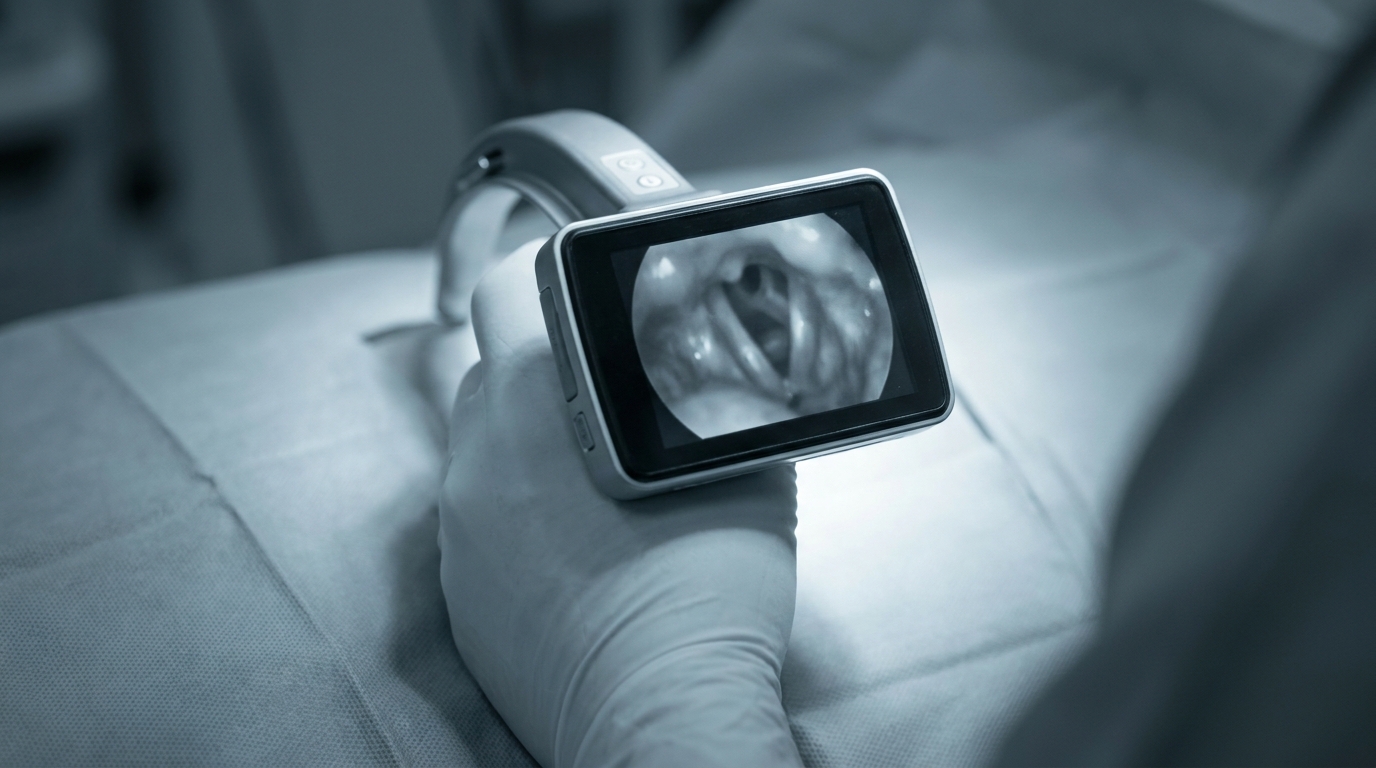
Endotracheal intubation is one of the most challenging procedures in emergency medicine – and simultaneously one where failure can immediately become life-threatening. Registry data consistently show that first-pass intubation success rates in prehospital and in-hospital emergency settings with conventional direct laryngoscopy (DL) can be below 80%. Each additional intubation attempt increases the risk of hypoxia, aspiration, esophageal intubation, and hemodynamic instability. Against this backdrop, video laryngoscopy (VL) has firmly established itself in airway management in recent years. Current evidence supports the use of video laryngoscopes as a first-line instrument, particularly in anticipated difficult airways – but even in seemingly straightforward intubations, you benefit from the improved view of the glottic plane. This article compares the most common systems, examines technical differences, and provides you with concrete tips for use under time pressure.
Why Video Laryngoscopy? – The Evidence
Several multicenter studies and meta-analyses demonstrate that video laryngoscopes significantly improve first-pass intubation success rates compared to direct laryngoscopy. The advantage is particularly pronounced in:
- Cormack-Lehane Grade III and IV on direct laryngoscopy
- Limited mouth opening (≥ 2 cm is sufficient for most VL blades)
- Limited cervical spine mobility (e.g., trauma with immobilization, ankylosing spondylitis)
- Obesity (BMI > 30, especially with a short neck)
- Blood, secretions, or vomitus in the pharynx – here the camera image often still provides orientation when direct vision is completely lost
At the same time, the literature shows that video laryngoscopes also improve success rates for providers with less intubation experience. This makes them particularly valuable in settings where intubation is not performed daily – such as in the resuscitation bay by non-anesthesiology personnel or in emergency medical services.
Honestly Acknowledging Limitations
VL is not a cure-all. An improved view of the glottis (better Cormack-Lehane grade) does not automatically correlate with successful tube placement. The so-called "video laryngoscopy paradox" describes exactly this situation: you see the vocal cords perfectly but cannot pass the tube through them. The causes usually lie in:
- Missing stylet or incorrectly pre-shaped stylet
- Too steep an insertion angle with hyperangulated blades
- Lack of practice in the specific tube guidance technique for the respective device
Blade Designs: Macintosh Geometry vs. Hyperangulated
Before we compare the specific devices, it is worth looking at the fundamental design philosophies:
Macintosh-Geometry Blades
- Blade profile resembles the classic Macintosh blade
- Allows both direct laryngoscopy (looking past the blade) and video-assisted intubation
- Tube guidance is similar to DL – a stylet is usually not mandatory
- Examples: C-MAC with Macintosh blade, McGrath MAC
Hyperangulated Blades
- Steeper curvature angle (usually 60–90°)
- Provide excellent visualization even in the most difficult anatomical conditions
- Mandatory use of a pre-shaped stylet (hockey stick shape, approximately 60°)
- Direct vision past the blade is virtually impossible by design
- Examples: C-MAC D-Blade, GlideScope (standard blade), McGrath X-Blade
Clinical Recommendation: In emergency medicine, a system that offers both blade geometries has proven effective. This way, you can switch from the Macintosh blade to the hyperangulated blade in case of an unexpectedly difficult airway without changing the entire device system.
Device Overview: C-MAC, GlideScope, McGrath
C-MAC (Karl Storz)
The C-MAC system by Karl Storz is widely used in German-speaking countries and has established itself in both in-hospital and prehospital settings.
Technical Features:
- Modular system: monitor (reusable) + blade (reusable or disposable)
- Available blades: Macintosh sizes 2–4, Miller 0–1, D-Blade (hyperangulated), C-MAC Pocket Monitor for mobile use
- Camera at the blade tip with LED illumination
- Integrated anti-fog system
- Recording function for documentation and teaching
Strengths:
- Most versatile blade selection on the market – from neonates (Miller 0) to morbidly obese adults (Macintosh 4 / D-Blade)
- Macintosh blade allows hybrid technique (direct + indirect laryngoscopy)
- Robust construction, clinically proven over many years
- Pocket Monitor variant is compact enough for the emergency bag
Weaknesses:
- Higher weight and larger form factor than compact disposable systems
- Reusable blades require reprocessing
- D-Blade has a steep learning curve for tube guidance
GlideScope (Verathon)
The GlideScope was one of the first commercially successful video laryngoscopes and has significantly shaped the concept of video laryngoscopy.
Technical Features:
- Available as GlideScope Titanium (reusable) and GlideScope Spectrum (disposable blade on reusable handle)
- Hyperangulated standard blade with 60° curvature
- Macintosh-style blade (available in newer generations)
- Monitor as a separate unit or tablet-based
- GlideStylet as a specially designed stylet included in delivery
Strengths:
- Excellent image quality with high resolution
- The specially designed GlideStylet significantly facilitates tube guidance with the hyperangulated blade
- Large body of evidence in the literature
- Spectrum series offers a hygienic disposable solution at reduced costs compared to fully disposable systems
Weaknesses:
- Standard hyperangulated blade requires consistent stylet use
- Separate monitor can be cumbersome in confined spaces (ambulance, helicopter)
- Higher acquisition cost compared to some competitors
McGrath MAC (Medtronic)
The McGrath MAC impresses with its compactness and is particularly popular in the prehospital setting.
Technical Features:
- Integrated system: handle with built-in LCD screen, snap-on disposable blade
- Blades: Macintosh geometry (sizes 2–4), McGrath X-Blade (hyperangulated, separate product)
- Battery-powered (approximately 250 minutes runtime)
- Weight approximately 175 g – extremely lightweight
Strengths:
- Lowest weight and most compact form factor of all common systems
- Monitor on the handle – no separate screen needed, no cable
- Disposable blades: no reprocessing, hygienic, rapidly deployable
- Macintosh geometry also allows direct laryngoscopy
- Intuitive handling, short learning curve
Weaknesses:
- Small integrated screen (2.5 inches) – with multiple team members, not everyone can see
- Disposable blades generate ongoing costs
- No neonatal blade in the standard range
- Image quality slightly below the larger systems
- Recording/documentation function not available as standard
Device Selection: Which System Fits Your Setting?
The choice of video laryngoscope depends on several factors. The following decision matrix helps with orientation:
| Criterion | C-MAC | GlideScope | McGrath MAC |
|---|---|---|---|
| Prehospital / EMS | ++ (Pocket Monitor) | + | +++ |
| Resuscitation Bay / Emergency Department | +++ | +++ | ++ |
| Anesthesia / OR | +++ | +++ | ++ |
| Pediatrics / Neonatology | +++ | + | – |
| Difficult Airway | +++ (D-Blade) | +++ | ++ (X-Blade) |
| Team Visibility | +++ (external monitor) | +++ | + (small screen) |
| Portability | + | + | +++ |
| Cost (Acquisition) | Medium–high | High | Low–medium |
| Cost (Consumables) | Low (reusable) | Medium | Medium (disposable blades) |
Pragmatic Advice: Many departments benefit from a dual-system approach – for example, McGrath MAC as the standard for rapid intubations and C-MAC with D-Blade as backup for the anticipated difficult airway.
Technique Under Time Pressure: 10 Practical Tips
1. Know Your Device
Practice handling your video laryngoscope before the emergency. This includes: powering on, mounting the blade, preparing the stylet, anti-fog measures. Under stress, unfamiliar devices cost valuable seconds.
2. Prepare the Stylet – Always
Even when using a Macintosh-geometry blade: have a pre-shaped stylet in the tube. With hyperangulated blades, it is mandatory. The ideal shape is the hockey stick – straight to the tube tip, then a 60° bend.
3. Optimize Positioning
The improved viewing angle gets you to the glottis quickly – but optimal patient positioning remains essential. Ramped position (upper body slightly elevated, tragus at sternal notch level) is the standard, especially in obese patients.
4. Do Not Insert Too Deep
A common beginner mistake: the blade is advanced too deep into the esophagus. All you see then is mucosa. Solution: Insert slowly and identify anatomical landmarks sequentially – uvula, epiglottis, arytenoid cartilages.
5. Eyes on the Monitor, Not in the Mouth
With hyperangulated blades, direct vision is not possible by design. But even with Macintosh-geometry VL, you should primarily look at the monitor. Switching between direct and indirect vision costs time and orientation.
6. Actively Guide the Tube
The tube does not automatically follow the blade curvature. Especially with hyperangulated blades, you must actively direct the tube tip toward the glottis. Technique: insert the tube from the right (to avoid obstructing the monitor view), steer the stylet tip toward the vocal cords, withdraw the stylet after passing through the glottis.
7. Don't Forget BURP/OELM
Even with a video laryngoscope, Backward-Upward-Rightward Pressure (BURP) or Optimal External Laryngeal Manipulation (OELM) can further improve the view. Communicate clearly with your team.
8. Have Suction Ready
Blood and secretions contaminate the camera lens and render the VL useless. Always keep a large-bore suction device (Yankauer or better: large-lumen suction catheter) within reach. In case of massive contamination: withdraw the blade, clean the lens, reinsert.
9. Cuff Manometry and Capnography
Successful tube placement is only confirmed after end-tidal CO₂ detection. The video image of vocal cord passage alone is not sufficient as the sole confirmation of placement – even if it appears convincing.
10. Define Plan B
Video laryngoscopy is an excellent tool, but not a substitute for a structured airway algorithm. Before each intubation attempt, define:
- Plan A: VL intubation (maximum 2 attempts)
- Plan B: Supraglottic airway device (laryngeal mask, i-gel)
- Plan C: Front-of-neck access (cricothyrotomy)
The current Difficult Airway Society (DAS) guidelines and AHA recommendations for airway management emphasize: a maximum of two intubation attempts in emergencies, then escalation. Each additional attempt worsens the prognosis.
Common Mistakes and How to Avoid Them
| Mistake | Consequence | Solution |
|---|---|---|
| No stylet with hyperangulated blade | Tube cannot reach the glottis | Always prepare a stylet |
| Blade inserted too deep | Esophageal view, loss of orientation | Insert slowly under direct visualization |
| Excessive levering force | Dental trauma, mucosal injury | VL requires less force than DL |
| Anti-fog measures neglected | Fogged lens, loss of vision | Use anti-fog wipe or pre-warm the blade |
| No capnography verification | Unrecognized esophageal intubation | etCO₂ after every intubation |
| No defined Plan B | Time lost on failure, "fixation error" | Establish airway algorithm in advance |
Integration into the Airway Algorithm
Video laryngoscopy does not replace the structured airway algorithm – it is one element within it. The AHA recommends in its current Advanced Cardiac Life Support (ACLS) guidelines that video laryngoscopy should be considered as the preferred technique when expertise is available. The key factor is team communication: who intubates, who assists, who suctions, who monitors the timer, who prepares Plan B.
In the resuscitation setting, an additional consideration applies: intubation must not cause an interruption of chest compressions for more than 10 seconds. Video laryngoscopy can help reduce intubation time – provided the operator is familiar with the device.
Summary of Key Messages
- Video laryngoscopy significantly improves first-pass intubation success rates and glottic visualization.
- The choice between Macintosh-geometry and hyperangulated blades depends on the clinical situation – ideally, you have access to both options.
- C-MAC, GlideScope, and McGrath MAC each have specific strengths. Device selection depends on the setting, patient population, and logistical conditions.
- Even with a perfect glottic view, tube placement can fail – correct stylet technique is essential.
- Maximum two intubation attempts, then escalation within the airway algorithm.
- Regular training on your specific device is the most important success factor.
Practical Training
Video laryngoscopy cannot be learned through reading alone. Handling the different blade geometries, tube guidance with a stylet, and structured management under time pressure require repeated hands-on practice – ideally in realistic simulation scenarios. In the Emergency Physician Refresher Course by Simulation Tirol, you train airway management on high-fidelity simulators, practice using common video laryngoscopes, and work through critical scenarios from the difficult airway to the can't-intubate-can't-oxygenate situation. This way, you solidify your skills under controlled conditions before they matter in the next real emergency.
More Articles
Acute Dyspnea: Differential Diagnosis and Immediate Management
Dyspnea has numerous causes ranging from asthma to heart failure to pulmonary embolism. This article presents a systematic approach to differentiation based on history, clinical examination, auscultation, and point-of-care diagnostics.
Acute Hypoglycemia: Emergency Management in Adults and Children
Threshold values, symptom recognition, oral glucose vs. IV dextrose vs. glucagon – with separate dosing for adult and pediatric patients. A common emergency presentation systematically reviewed.
Acute Adrenal Insufficiency: Addisonian Crisis in the Emergency Setting
Adrenal crisis is frequently misdiagnosed and can be fatal. This article describes at-risk patients, clinical signs, immediate hydrocortisone administration, and the management of accompanying hypoglycemia and hyperkalemia.