Pulmonary Edema in Emergency Medicine: Symptoms, Causes, and Treatment
Acute pulmonary edema is a common emergency with high mortality. This article covers the differentiation of cardiogenic vs. non-cardiogenic pulmonary edema, immediate interventions such as NIV, loop diuretics, and nitrates, as well as guideline-based escalation strategies.

Author: Dr. med. univ. Daniel Pehböck, DESA
Specialist in Anesthesiology and Intensive Care Medicine, AHA-certified ACLS/PALS Instructor, Course Director Simulation Tirol
Reading time approx. 8 min
Acute pulmonary edema is one of the most time-critical emergencies in both in-hospital and prehospital care. Fluid accumulation in the pulmonary interstitium and alveoli leads to rapidly progressive respiratory failure that, without targeted therapy, can result in respiratory arrest and circulatory collapse within a short time. Mortality of cardiogenic pulmonary edema ranges from 10–20% during the initial hospital admission depending on the study—and is significantly higher when accompanied by cardiogenic shock. What is decisive for outcome is that you rapidly differentiate the cause, initiate acute therapy in parallel, and follow a clear escalation plan. This article provides you with the pathophysiological fundamentals, clinical differentiation, and a structured treatment algorithm based on current evidence.
Pathophysiology: Why the Lungs Are "Underwater"
The balance between hydrostatic pressure, oncotic pressure, and alveolocapillary membrane integrity determines the fluid homeostasis of the lungs. A disturbance in any of these systems leads to transudation or exudation of fluid into the interstitium and ultimately into the alveoli.
Cardiogenic Pulmonary Edema
In cardiogenic pulmonary edema, the pulmonary capillary wedge pressure (PCWP) rises above the critical threshold of approximately 18 mmHg. The most common causes:
- Acute left heart failure (systolic or diastolic)
- Acute coronary syndrome with pump failure
- Acute mitral valve regurgitation (e.g., papillary muscle rupture, chordae tendineae rupture)
- Hypertensive crisis (flash pulmonary edema)
- Tachycardic or bradycardic arrhythmias with hemodynamic compromise
- Acute decompensation of chronic heart failure (e.g., due to infection, non-compliance, volume overload)
Increased preload and/or afterload leads to congestion in the pulmonary circulation. The initial interstitial fluid accumulation (interstitial edema) progresses to alveolar edema with further pressure increases. Clinically, this correlates with the transition from dyspnea and tachypnea to the classic presentation with frothy, sometimes pink-tinged sputum.
Non-Cardiogenic Pulmonary Edema
Here, the alveolocapillary membrane is directly damaged, leading to increased permeability. The PCWP typically remains normal. Important causes:
- ARDS (Acute Respiratory Distress Syndrome) – due to pneumonia, sepsis, aspiration, trauma
- Inhalation injury (toxic gases, smoke inhalation)
- High-altitude pulmonary edema (HAPE)
- Neurogenic pulmonary edema (after traumatic brain injury, subarachnoid hemorrhage)
- Negative-pressure pulmonary edema (after airway obstruction, e.g., laryngospasm)
- Re-expansion pulmonary edema (after drainage of a pneumothorax or large pleural effusions)
- Drug-induced (e.g., opioids, salicylates)
The differentiation is of central therapeutic importance: while cardiogenic pulmonary edema focuses on preload and afterload reduction, non-cardiogenic pulmonary edema requires cause-specific therapy and a lung-protective ventilation strategy.
Clinical Presentation and Diagnostics
Cardinal Symptoms
The clinical picture is often striking and can usually be identified quickly:
- Severe dyspnea with orthopnea (the patient sits upright, bracing themselves)
- Tachypnea (respiratory rate often > 30/min)
- Use of accessory respiratory muscles, jugular retractions
- Bilateral wet crackles, often audible up to the lung apices
- Frothy, sometimes pink-tinged sputum
- Tachycardia, often with diaphoresis
- Cyanosis in advanced stages
- Anxiety and agitation as an expression of respiratory distress
Diagnostic Algorithm
Parallel to immediate therapy, you should initiate a structured diagnostic workup:
Basic diagnostics:
- SpO₂ monitoring – initially often < 90%
- 12-lead ECG – signs of ischemia? Arrhythmia? Evidence of right heart strain?
- Blood pressure – hypertensive pulmonary edema vs. cardiogenic shock (systolic < 90 mmHg)
- History – cardiac history, medications, triggers
Advanced diagnostics:
- Point-of-care ultrasound (POCUS): This has revolutionized the diagnosis of pulmonary edema. Bilateral multiple B-lines (≥ 3 per intercostal space) indicate alveolar edema. Cardiac assessment provides information about LV function, valvular pathology, and pericardial effusion. Assessment of the inferior vena cava provides clues about volume status.
- Arterial blood gas analysis (ABG): Typical findings include hypoxemic respiratory failure, initially often with respiratory alkalosis (hyperventilation), progressing to respiratory acidosis with exhaustion. Concomitant metabolic acidosis (elevated lactate) is a warning sign of organ hypoperfusion.
- Laboratory diagnostics: BNP/NT-proBNP (elevated in cardiogenic pulmonary edema, differentiation at borderline values), troponin, creatinine, electrolytes, complete blood count, CRP/PCT if infection is suspected.
- Chest X-ray: Classic signs of cardiogenic pulmonary edema include Kerley B lines, peribronchial cuffing, cephalization of pulmonary vessels, butterfly-shaped perihilar opacification, and pleural effusions. However, sensitivity is lower than that of POCUS, and the chest X-ray cannot replace therapy in the acute phase.
Differentiation: Cardiogenic vs. Non-Cardiogenic
| Feature | Cardiogenic | Non-cardiogenic |
|---|---|---|
| History | Heart failure, CAD, valvular disease | Sepsis, pneumonia, trauma, inhalation |
| BNP/NT-proBNP | Markedly elevated | Normal to mildly elevated |
| PCWP (invasive) | > 18 mmHg | ≤ 18 mmHg |
| Echocardiography | LV dysfunction, valvular pathology | Usually unremarkable |
| Chest X-ray | Cardiac congestion pattern, effusions | Diffuse bilateral infiltrates |
| Response to diuretics/nitrates | Good | Poor |
In clinical reality, mixed presentations exist—for example, in a septic patient with pre-existing heart failure. Here, the overall picture from clinical findings, laboratory results, and imaging helps guide decision-making.
Immediate Treatment of Acute Cardiogenic Pulmonary Edema
Therapy follows the principle: Secure oxygenation – reduce preload – reduce afterload – treat the cause. Critical is the classification by blood pressure level, as this determines the pharmacological strategy.
Positioning and General Measures
- Upright positioning (sitting position with legs hanging down) – reduces preload and improves respiratory mechanics
- Reassurance of the patient – anxiety increases sympathetic activation and oxygen consumption
- Intravenous access – at least one large-bore IV line
- Monitoring: SpO₂, ECG, blood pressure (frequent, ideally every 3–5 minutes)
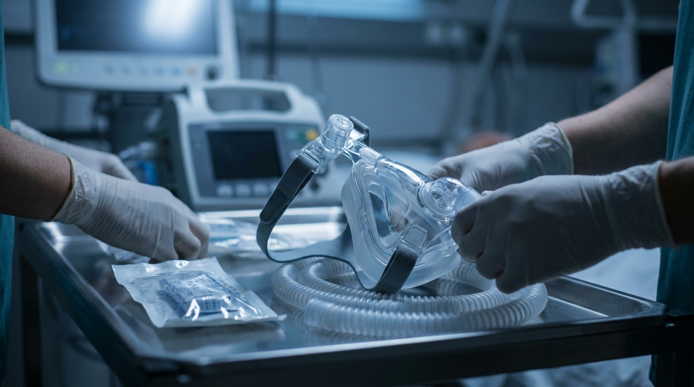
Non-Invasive Ventilation (NIV)
NIV is the intervention with the strongest evidence in the acute treatment of cardiogenic pulmonary edema. It has been shown to reduce both intubation rates and mortality.
- Mode: CPAP (Continuous Positive Airway Pressure) or BiPAP/BiLevel (preferred in hypercapnia)
- PEEP/EPAP: Start at 5–8 cmH₂O, titrate to 10–12 cmH₂O based on tolerance and effect
- FiO₂: Adjust to SpO₂ target (92–96%)
- Mechanism of action: Positive end-expiratory pressure recruits collapsed alveoli, reduces intrapulmonary shunt, lowers preload (decreased venous return), and reduces afterload (unloading the left ventricle by reducing transmural pressure)
Caution: In patients with cardiogenic shock (systolic blood pressure < 90 mmHg), NIV may further compromise circulation through preload reduction. Close monitoring or primary intubation should be considered here.
Pharmacological Therapy by Blood Pressure Level
Hypertensive Pulmonary Edema (Systolic BP > 140 mmHg)
This is the most common presentation and has the best prognosis. The focus is on aggressive afterload reduction:
- Glyceryl trinitrate (nitroglycerin):
- Sublingual: 0.4–0.8 mg, repeatable every 3–5 minutes
- Intravenous (preferred in severe symptoms): Start at 10–20 µg/min, titrate in 10–20 µg increments every 3–5 minutes, target dose often 100–200 µg/min
- Acts primarily as a venous dilator (preload reduction), at higher doses also arterial (afterload reduction)
- Urapidil: 10–50 mg slow IV bolus, well titratable, particularly useful in pronounced hypertension
Caution: Nitrates are contraindicated with concurrent use of PDE-5 inhibitors (e.g., sildenafil), in aortic stenosis, and in right ventricular infarction.
Normotensive Pulmonary Edema (Systolic BP 100–140 mmHg)
- Glyceryl trinitrate cautiously titrated (IV preferred)
- Loop diuretics:
- Furosemide: 20–40 mg IV in diuretic-naive patients; in patients on chronic diuretic therapy, at least the oral daily dose as an IV bolus
- Effect: Acute venous vasodilation (minutes before the diuretic effect), followed by diuresis
- Current evidence shows that while diuretics are symptomatically effective, they should be used conservatively in the absence of volume overload. In hypertensive flash pulmonary edema, afterload reduction is more important than diuresis.
Hypotensive Pulmonary Edema / Cardiogenic Shock (Systolic BP < 90 mmHg)
This is the prognostically most unfavorable constellation. Nitrates and high-dose diuretics are contraindicated here:
- Vasopressors:
- Norepinephrine: 0.1–1 µg/kg/min – agent of choice for stabilizing MAP (target ≥ 65 mmHg)
- Inotropes:
- Dobutamine: 2–20 µg/kg/min – for confirmed severe LV dysfunction with low cardiac output
- Levosimendan: 0.05–0.2 µg/kg/min – calcium sensitizer, particularly useful in beta-blocker-induced low cardiac output
- Milrinone: 0.375–0.75 µg/kg/min – PDE-3 inhibitor, caution with hypotension
- Mechanical circulatory support: In refractory cardiogenic shock, consider IABP (intra-aortic balloon pump), Impella, or VA-ECMO at specialized centers
- Intubation and mechanical ventilation: In treatment-refractory hypoxemia, exhaustion, or altered level of consciousness
Morphine – A Critical Look
Morphine was traditionally used as a standard component of pulmonary edema therapy (anxiolysis, preload reduction). However, current data show that morphine is associated with increased intubation rates and potentially increased mortality. It should therefore not be used routinely. If at all, then at the lowest dose (e.g., 1–2 mg IV fractionated) for severe agitation not manageable by other means—and with close monitoring of respiratory rate.
Cause-Specific Therapy
Stabilization alone is not enough—you must identify and treat the underlying cause:
- ACS: Emergency coronary angiography for STEMI, early invasive strategy for NSTEMI with hemodynamic instability
- Hypertensive crisis: Controlled blood pressure reduction (target: 25% reduction in the first hour)
- Tachyarrhythmia: Cardioversion (electrical or pharmacological, depending on stability)
- Bradycardia: Atropine, transcutaneous pacing
- Acute valvular regurgitation: Emergency surgical intervention
- Volume overload in dialysis patients: Emergency dialysis
Treatment of Non-Cardiogenic Pulmonary Edema
In non-cardiogenic pulmonary edema, causal therapy takes priority:
- ARDS: Lung-protective ventilation (tidal volume 6 mL/kg ideal body weight, plateau pressure < 30 cmH₂O, adequate PEEP), prone positioning in moderate/severe ARDS, restrictive fluid strategy
- Sepsis: Source control, empiric antibiotic therapy, hemodynamic stabilization
- Inhalation injury: Bronchoscopy, supportive ventilation, antidote administration for specific toxins
- High-altitude pulmonary edema: Descent, supplemental oxygen, nifedipine, portable hyperbaric chamber if needed
Common Mistakes and Pitfalls
- Overly aggressive diuresis in flash pulmonary edema: These patients are often euvolemic—afterload reduction is key, not volume removal.
- Routine morphine administration: Obsolete due to associated risks.
- Delayed initiation of NIV: The earlier NIV is established, the better the outcome. Don't wait for the chest X-ray.
- Failure to search for the underlying cause: Symptom control is important, but without identifying and treating the trigger, recompensation is likely to fail.
- Overlooking cardiogenic shock: Blood pressure must be monitored closely. An initially normotensive patient can rapidly decompensate—especially under nitrate therapy.
Summary: Structured Action Algorithm
- Recognition – Dyspnea, orthopnea, wet crackles, SpO₂ ↓
- Positioning – Upper body elevated, legs dependent
- NIV – CPAP/BiLevel, PEEP 5–10 cmH₂O, initiate early
- Blood pressure-guided pharmacotherapy:
- Hypertensive → Aggressive nitrates, moderate diuretics
- Normotensive → Cautious nitrates, diuretics
- Hypotensive → Vasopressors, inotropes, no nitrates/diuretics
- Diagnostics in parallel: POCUS, ECG, ABG, laboratory
- Treat the cause: ACS → catheterization, arrhythmia → conversion, volume overload → diuresis/dialysis
- Escalation: Intubation if NIV fails, mechanical circulatory support in refractory shock
Practical Training
Acute pulmonary edema demands structured action under time pressure—from clinical assessment through blood pressure-adapted pharmacotherapy to escalation decisions. These skills are best trained in realistic simulation scenarios. In the ACLS course by Simulation Tirol (AHA-certified), you practice exactly these situations: systematic patient assessment, pharmacological algorithms, and team communication in cardiovascular emergency care. The combination of theory and hands-on simulator training provides the confidence you need when it matters most.
Want to practice this hands-on?
In our ACLS-Kurs (Advanced Cardiac Life Support) you practice this topic hands-on with high-tech simulators and experienced instructors.
More Articles
Acute Dyspnea: Differential Diagnosis and Immediate Management
Dyspnea has numerous causes ranging from asthma to heart failure to pulmonary embolism. This article presents a systematic approach to differentiation based on history, clinical examination, auscultation, and point-of-care diagnostics.
Acute Hypoglycemia: Emergency Management in Adults and Children
Threshold values, symptom recognition, oral glucose vs. IV dextrose vs. glucagon – with separate dosing for adult and pediatric patients. A common emergency presentation systematically reviewed.
Acute Adrenal Insufficiency: Addisonian Crisis in the Emergency Setting
Adrenal crisis is frequently misdiagnosed and can be fatal. This article describes at-risk patients, clinical signs, immediate hydrocortisone administration, and the management of accompanying hypoglycemia and hyperkalemia.