Cricothyrotomy: Indications, Technique, and the Surgical Airway
Cricothyrotomy is the last resort in a cannot-intubate-cannot-oxygenate scenario. This article covers indications, surgical vs. needle technique, anatomical landmarks, and pediatric considerations.

Author: Dr. med. univ. Daniel Pehböck, DESA
Specialist in Anesthesiology and Intensive Care Medicine, AHA-certified ACLS/PALS Instructor, Course Director Simulation Tirol
Reading time approx. 8 min
Cricothyrotomy is the most invasive yet most definitive step in airway management – and at the same time one of the rarest procedures in clinical practice. That's precisely the problem: when you need it, you need it immediately, but very few emergency physicians and anesthesiologists have ever performed one on a living patient. The cannot-intubate-cannot-oxygenate (CICO) scenario is one of the few situations in medicine where seconds literally determine life or death. If you hesitate in that moment, you lose precious time. If you're prepared, you can save a life. This article provides you with the anatomical fundamentals, indications, surgical and needle techniques, as well as pediatric considerations – evidence-based, practical, and thorough.
The CICO Scenario: When Does Cricothyrotomy Become Necessary?
Cricothyrotomy stands at the end of all established airway algorithms. The AHA guidelines, DAS guidelines (Difficult Airway Society), and DGAI recommendations all follow a stepwise approach to difficult airway management:
- Optimized laryngoscopy (including video laryngoscopy, bougie, positioning)
- Supraglottic airway device (laryngeal mask, i-gel, laryngeal tube)
- Return to bag-mask ventilation (two-hand technique, oropharyngeal airway)
- Surgical airway – cricothyrotomy
The indication is clearly defined: you can neither intubate nor oxygenate the patient, oxygen saturation is dropping, and all less invasive measures have failed. The critical question is not "if" but "when" you take the step to cricothyrotomy. Current evidence clearly shows: the most common source of error is not technical failure of the cricothyrotomy itself, but delayed decision-making. Repeated futile intubation attempts delay the surgical airway and lead to prolonged hypoxia.
Typical Clinical Scenarios
- Massive facial or neck trauma with bleeding and swelling of the upper airway
- Anaphylaxis with complete glottic edema despite epinephrine administration
- Burns/inhalation injury with progressive airway swelling
- Tumors or abscesses in the pharyngeal/laryngeal region
- Postoperative airway obstruction following thyroid or ENT surgery (hematoma)
- Trismus (e.g., tetanus, abscess, post-radiation)
Anatomical Landmarks: The Key to Success
Anterior neck anatomy is the foundation of every successful cricothyrotomy. Without reliable identification of landmarks, the risk of complications increases substantially.
The Cricothyroid Ligament (Ligamentum Conicum)
The target structure is the cricothyroid membrane – the fibroelastic membrane between the inferior border of the thyroid cartilage and the superior border of the cricoid cartilage. This membrane is:
- Approximately 9–10 mm in height and 22–30 mm in width
- Relatively avascular in the midline
- Covered only by skin, subcutaneous tissue, and superficial fascia
Palpation – Step by Step
- Identify the laryngeal prominence (Adam's apple) – usually easily palpable in men, more difficult in women and obese patients.
- Slide the index finger caudally – the first palpable depression below the thyroid cartilage is the cricothyroid membrane.
- Palpate the cricoid cartilage – the firm caudal resistance below the membrane confirms correct localization.
A critical clinical point: in up to 30% of patients – especially obese women, those with short necks, or neck edema – palpation of the landmarks is considerably more difficult. Ultrasound-guided identification of the cricothyroid membrane prior to planned airway management in anticipated difficult airways is therefore gaining increasing importance. This preparation – known as "airway mapping" – can be documented in advance using a skin marker.
Vascular Supply and Risk Zones
The cricothyroid artery (a branch of the superior thyroid artery) typically runs in the upper third of the membrane, often horizontally. A midline incision in the lower to middle third of the membrane minimizes bleeding risk. Unusual vascular variants – including a high-riding brachiocephalic trunk – are rare but have been described.
Surgical Cricothyrotomy: The Method of Choice
Current evidence and recommendations from most professional societies clearly favor the surgical (open) cricothyrotomy as the method of choice in emergencies. The surgical technique has a higher first-attempt success rate and a lower complication rate compared to the needle technique.
Required Equipment
- Scalpel (blade No. 10 or No. 20 – broad blade preferred)
- Bougie (Eschmann introducer) or alternatively a dedicated tracheal tube introducer
- Tracheal tube size 6.0 (inner diameter) – cuffed
- Alternatively: dedicated cricothyrotomy cannula (e.g., cuffed cricothyrotomy tube)
- Clamp or Kilner hook (helpful but not mandatory)
- Suction
Technique: The Bougie-Guided Horizontal Incision
The best-validated and currently recommended technique is the "scalpel-bougie-tube" method. It follows a clear, standardized sequence:
Step 1 – Skin Incision: Vertical skin incision (approximately 4–5 cm in length) over the estimated location of the cricothyroid membrane. The vertical incision has the advantage of allowing adequate exploration even when landmark identification is uncertain and reduces the likelihood of lateral vessel injury.
Step 2 – Membrane Identification: Blunt dissection of the subcutaneous tissue with the fingers until the membrane is palpable and visible. In slim patients, this step is often minimal.
Step 3 – Membrane Incision: Horizontal stab incision through the cricothyroid membrane in the lower third of the membrane. The scalpel is stabilized to prevent excessive depth of penetration and injury to the posterior tracheal wall. Tip: leave the scalpel in place initially after the incision and use it as a placeholder so you don't lose the opening.
Step 4 – Bougie Insertion: Insert a bougie with the coudé tip directed caudally (toward the trachea) through the incision. Palpate tracheal rings as tactile feedback – "clicking" confirms correct intratracheal placement.
Step 5 – Tube Advancement: Advance a tracheal tube (ID 6.0) over the bougie into the trachea. Inflate the cuff, initiate ventilation, and confirm placement with capnography.
Pitfalls and Practical Tips
- Too timid an incision: A superficial incision that does not penetrate the entire membrane is a common error. The membrane is tough – a decisive cut is necessary.
- Loss of landmarks: After the skin incision, orientation can be lost, especially in bloody conditions. Therefore: stabilize the larynx with your non-dominant hand and do not let go (the so-called "laryngeal handshake").
- Wrong level: An incision through the membrane between the cricoid cartilage and the first tracheal ring, or even further caudally (tracheotomy location), is an avoidable complication. Consistent palpation before the incision is critical.
- Tube too large: A tube with an inner diameter greater than 6.0–6.5 mm often will not fit through the cricothyrotomy opening and causes trauma or malpositioning.
Needle Cricothyrotomy: When and for Whom?
With the needle technique, the cricothyroid membrane is punctured with a cannula (e.g., Seldinger technique or dedicated kits such as Melker, Quicktrach), creating a narrow-bore access. Commercial kits are available in various designs.
Advantages
- Technically easier to learn
- Less initial tissue damage
- Less bleeding
Disadvantages
- Higher failure rate compared to the surgical technique
- Narrow-bore cannulae do not allow adequate ventilation against high airway resistance
- Kinking and dislocation of the cannula are common problems
- In adults, adequate oxygenation and CO₂ elimination through small cannulae (≤ 4 mm ID) is virtually impossible – jet ventilation would be required, but carries a significant risk of barotrauma
The current recommendation is therefore: For adults, surgical cricothyrotomy is the method of choice. The needle technique may serve as a bridging measure but should not be considered a definitive airway.
Pediatric Considerations
Cricothyrotomy in children poses particular challenges and differs from the adult approach in several important ways.
Anatomical Differences
- The larynx is positioned higher (at the level of C3–C4 in infants vs. C5–C6 in adults)
- The cricothyroid membrane is significantly smaller – only approximately 2.5–3 mm in height in infants
- The cricoid cartilage is the narrowest point of the pediatric airway and the only completely closed cartilaginous structure – injury can lead to subglottic stenosis
- Landmarks are more difficult to palpate
Recommended Approach by Age
- Children under 8–10 years: Surgical cricothyrotomy is considered relatively contraindicated by many professional societies. Instead, needle cricothyrotomy with a large-bore cannula (14G or 16G) and jet ventilation or bag-cannula ventilation is recommended as a bridging measure until definitive surgical tracheotomy.
- Children approximately 10–12 years and older: Surgical cricothyrotomy can be performed analogously to the adult approach, but with a smaller tube (ID 4.0–5.0).
Needle Cricothyrotomy – Technique in Children
- Identify the cricothyroid membrane (ultrasound-guided if possible)
- Puncture with a 14G cannula attached to a syringe (filled with saline) at a 45° angle directed caudally
- Aspiration of air confirms intratracheal placement
- Advance the plastic cannula, remove the needle
- Connect to a bag-valve device via a 3.0 tube connector or dedicated adapter
- Jet ventilation: lowest possible pressure, I:E ratio approximately 1:4, always ensure adequate expiration (caution: barotrauma!)
Key point: Needle cricothyrotomy in children is a temporary emergency solution, not a definitive airway. It buys time to organize a surgical tracheotomy under controlled conditions.
Complications and Their Management
Like any invasive procedure, cricothyrotomy carries risks. The most important complications are:
- Malplacement (pretracheal, esophageal, paramedian): capnography is the gold standard for confirming placement
- Bleeding: Usually from injury to the cricothyroid artery or pre-/paratracheal veins – typically manageable with compression
- Posterior tracheal wall injury: From cutting too deep or forcing the tube during insertion
- Subglottic stenosis: Especially in children and with prolonged cannulation – the cricothyrotomy must be converted to a formal tracheotomy within 24–72 hours
- Pneumomediastinum / subcutaneous emphysema: Particularly with jet ventilation via needle cricothyrotomy
Preparation and Team Aspects
Cricothyrotomy is not just a manual skill but also a team effort. The following aspects have been shown to improve success rates:
- Clear role assignment before every planned airway management in anticipated difficult airways: who will perform the cricothyrotomy if it becomes necessary?
- Equipment preparation: A pre-assembled cricothyrotomy kit (scalpel, bougie, tube 6.0) should be readily available for every difficult airway management
- Verbal declaration: The explicit announcement "CICO – we are now performing a cricothyrotomy" is essential to focus the team and break cognitive fixation on further intubation attempts
- Human factors: The psychological barrier to cutting open a patient's neck is enormous. Only regular training on models and simulators can lower this threshold sufficiently to enable decisive action in a real emergency
Summary: Key Points
- Cricothyrotomy is the last resort in a CICO scenario – and must not be indicated too late
- Anatomical landmarks (thyroid cartilage, cricothyroid membrane, cricoid cartilage) must be reliably identified – ultrasound-guided airway mapping in advance if needed
- The surgical "scalpel-bougie-tube" method is the recommended technique in adults
- In children under 8–10 years, needle cricothyrotomy with bridging ventilation is preferred over the surgical approach
- Capnography is mandatory for confirming placement
- Conversion to a tracheotomy within 24–72 hours
- Regular training is the only way to maintain the manual skills and decision-making competence for this rare but life-saving procedure
Practical Training
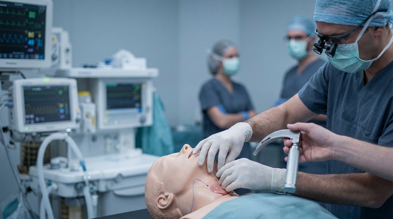
Cricothyrotomy is one of those procedures you should never perform for the first time on an actual patient – yet it occurs so rarely in clinical practice that building practical experience is nearly impossible. This makes regular, realistic simulation training all the more important. In the Emergency Physician Refresher Course by Simulation Tirol, you have the opportunity to work through the CICO scenario in a structured format: from recognizing the difficult airway, through establishing the indication, to hands-on performance of the surgical cricothyrotomy on a model. Together with experienced instructors, you train not only the technical skills but also team communication and decision-making under stress. You can find all course details at simulation.tirol/kurse/notarzt-refresher.
More Articles
Acute Dyspnea: Differential Diagnosis and Immediate Management
Dyspnea has numerous causes ranging from asthma to heart failure to pulmonary embolism. This article presents a systematic approach to differentiation based on history, clinical examination, auscultation, and point-of-care diagnostics.
Acute Hypoglycemia: Emergency Management in Adults and Children
Threshold values, symptom recognition, oral glucose vs. IV dextrose vs. glucagon – with separate dosing for adult and pediatric patients. A common emergency presentation systematically reviewed.
Acute Adrenal Insufficiency: Addisonian Crisis in the Emergency Setting
Adrenal crisis is frequently misdiagnosed and can be fatal. This article describes at-risk patients, clinical signs, immediate hydrocortisone administration, and the management of accompanying hypoglycemia and hyperkalemia.