Emergency Airway Management: Algorithm and Techniques
From head-tilt chin-lift to laryngeal mask to emergency cricothyrotomy – this article provides a structured overview of the airway algorithm including videolaryngoscopy, bougie technique, and the cannot-intubate-cannot-oxygenate scenario.

Author: Dr. med. univ. Daniel Pehböck, DESA
Specialist in Anesthesiology and Intensive Care Medicine, AHA-certified ACLS/PALS Instructor, Course Director Simulation Tirol
Reading time approx. 9 min
Airway management is one of the most critical competencies in emergency medicine. A lost airway means a lost patient – this simple truth makes structured mastery of the airway algorithm a matter of survival. It's not just about intubation itself, but about a stepwise approach from simple basic measures to surgical airway management. Especially under stress, time pressure, and adverse conditions – whether prehospital or in the emergency department – a clearly internalized algorithm determines success or failure. This article provides you with a structured walkthrough of all escalation levels, from head-tilt chin-lift to emergency cricothyrotomy.
Fundamental Principle: The Stepwise Airway Algorithm
Every airway algorithm follows an escalating stepwise principle. You start with the simplest measure and only advance to the next level when the previous one fails. One overriding goal applies: Oxygenation takes priority over intubation. Adequate bag-mask ventilation (BMV) is always more valuable than a desperate, repeated intubation attempt.
The escalation levels at a glance:
- Basic measures – Opening and maintaining the airway
- Bag-mask ventilation – Oxygenation and ventilation
- Supraglottic airway devices – Laryngeal mask, laryngeal tube
- Endotracheal intubation – Videolaryngoscopy as the standard
- Surgical airway – Cricothyrotomy as the last resort
Crucially, you must stay time-limited at each level. Two failed intubation attempts should lead you to a supraglottic alternative, not to a third attempt. Each attempt prolongs apnea time, worsens visualization due to edema and bleeding, and increases the risk of aspiration.
Basic Measures: The Underestimated First Step
Before you reach for any device, manual basic maneuvers come first. They are simple, but their importance can hardly be overstated.
Head-Tilt Chin-Lift
The standard maneuver for unconscious patients without suspected trauma. Extension of the cervical spine lifts the base of the tongue off the posterior pharyngeal wall and opens the airway. At the same time, you lift the chin to displace the mandible anteriorly.
Jaw-Thrust Maneuver
When cervical spine trauma is suspected, the jaw-thrust maneuver is the method of choice. You grasp both angles of the mandible and push it anteriorly without moving the cervical spine. In combination with manual in-line stabilization (MILS), this is the standard for trauma patients.
Suction Readiness
Before you perform any airway intervention, a functioning suction device must be at the ready. Blood, secretions, and vomitus in the upper airway are the most common preventable cause of failed laryngoscopy. A large-bore, rigid suction catheter (Yankauer-type) should be within arm's reach.
Oropharyngeal Airway (Guedel) and Nasopharyngeal Airway (NPA)
These simple adjuncts keep the airway open and significantly facilitate BMV. The Guedel airway is used in unconscious patients without a gag reflex. The nasopharyngeal airway is often better tolerated and can also be used in patients with reduced consciousness – use caution in the presence of midface fractures and coagulopathies.
Bag-Mask Ventilation: The Bridge to Definitive Airway Management
BMV is not a stopgap solution but the central technique for oxygenation. It must be mastered reliably, as it is your fallback at every level of the algorithm.
Optimizing BMV
If mask ventilation is unsuccessful, the acronym BONES helps identify risk factors:
- Beard – Impaired mask seal
- Obesity – Increased ventilation pressures required
- No teeth – Missing teeth worsen the mask fit
- Elderly – Reduced muscle tone
- Snoring/Sleep Apnea – Habitual airway obstruction
Practical measures for difficult BMV:
- Two-person technique – One person holds the mask bimanually with a double C-E grip, the other squeezes the bag
- Insert a Guedel or nasopharyngeal airway
- Optimize head position – Head-up positioning (ramping) for obese patients
- Reduce pressure – Slow, steady insufflation over 1 second to avoid gastric insufflation
Supraglottic Airway Devices: The Underestimated Escalation Level
Supraglottic airway devices (SGAs) have evolved from a last resort to an integral part of the airway algorithm. They should be viewed as Plan B for failed intubation and as Plan A for certain indications.
Laryngeal Mask
Second-generation laryngeal masks (e.g., i-gel, LMA Supreme, Ambu AuraGain) offer not only ventilation but also a gastric drainage channel and allow fiberoptic intubation through the device. Size selection is weight-based.
Laryngeal Tube
The laryngeal tube is particularly common in the prehospital setting and can be safely placed even by less experienced personnel. It provides a reliable airway but has limitations regarding secondary intubation.
When to Use SGA Instead of Intubation?
- Resuscitation by personnel not experienced in intubation
- Difficult airway after a failed first intubation attempt
- As a bridge device for further planning
- When oxygenation via SGA is successful: No reason to intubate under time pressure
Endotracheal Intubation: Videolaryngoscopy as the Standard
Current evidence clearly supports videolaryngoscopy (VL) as the first-line technique in emergency situations. VL significantly improves the first-pass intubation success rate compared to direct laryngoscopy, especially in difficult airways and with less experienced operators.
Rapid Sequence Induction (RSI)
RSI is the standard for emergency intubation in non-fasted patients. The procedure in brief:
- Preoxygenation – 3–5 minutes with 100% O₂ via a tight-fitting mask, target SpO₂ ≥ 95% (ideally supplemented via high-flow nasal cannula for apneic oxygenation)
- Apneic oxygenation – Nasal cannula at 15 L/min O₂ left in place during laryngoscopy
- Induction – e.g., ketamine 1.5–2 mg/kg IV or propofol 1.5–2.5 mg/kg IV (hemodynamically adapted)
- Neuromuscular blockade – Rocuronium 1.2 mg/kg IV (full intubating dose) or succinylcholine 1–1.5 mg/kg IV
- Intubation – After onset of paralysis (succinylcholine ~45–60 seconds, rocuronium ~60–90 seconds)
- Confirmation of placement – Capnography (end-tidal CO₂) as the gold standard
Videolaryngoscopy: Practical Tips
- Blade selection: Hyperangulated blades (e.g., D-Blade, McGrath MAC) are advantageous for anticipated difficult airways. Macintosh-style video blades allow both direct and indirect visualization.
- Pre-shape the tube with a stylet: With hyperangulated blades, a hockey stick–shaped stylet is mandatory.
- Bougie technique: The bougie (elastic introducer) is a game-changer. The current recommendation is to use the bougie routinely on the first intubation attempt – not just on the second. Technique: Advance the bougie under direct vision into the trachea, feeling for the characteristic "clicks" over the tracheal rings as tactile confirmation, then railroad the tube over the bougie.
- BURP/ELM maneuver: Backward-Upward-Rightward Pressure on the thyroid cartilage can improve the view of the glottis. External Laryngeal Manipulation (ELM) is directed by the intubator themselves and is preferred over blind BURP.
Cormack-Lehane Classification
The view of the glottis is documented in a standardized fashion:
- Grade I – Vocal cords fully visible
- Grade II – Only the posterior portion of the vocal cords visible (IIa) or only the arytenoid cartilages (IIb)
- Grade III – Only the epiglottis visible
- Grade IV – No glottic structures visible
From Grade IIb, intubation difficulty increases significantly. This is where you benefit most from bougie technique and a hyperangulated videolaryngoscope.
Maximum Number of Attempts
The recommendation is clear: A maximum of two intubation attempts before switching to an alternative strategy. Each attempt should last no longer than 30 seconds. Between attempts, reoxygenation is performed (BMV or SGA). A third attempt should only be made by a more experienced operator and/or with a different technique.
Cannot Intubate, Cannot Oxygenate (CICO): Emergency Cricothyrotomy
The CICO scenario is rare (estimated at 1:50,000 anesthetics), but when it occurs, every second counts. The diagnosis is: Intubation has failed AND oxygenation via BMV and SGA has failed. Now a surgical airway is the only option.
Recognizing the CICO Scenario
The greatest barrier is cognitive: Recognizing and accepting that you are in a CICO scenario. Fixation error – repeatedly attempting the same failed technique – is the most common cause of delays. Clear announcements within the team help: "We are in a cannot-intubate-cannot-oxygenate situation. I am now performing a cricothyrotomy."
Emergency Cricothyrotomy Technique
The surgical cricothyrotomy (scalpel-bougie-tube technique) is recommended as the standard approach:
- Identify landmarks – Palpate the cricothyroid membrane between the thyroid and cricoid cartilages. In obese patients or with difficult landmarks: Laryngeal handshake (bimanual grasp of the larynx, sliding down to the cricothyroid membrane)
- Skin incision – Horizontal incision over the cricothyroid membrane, approximately 3–4 cm, through skin and subcutaneous tissue
- Membrane incision – Horizontal incision of the cricothyroid membrane (stab-rotation technique: insert the scalpel horizontally, rotate 90° to enlarge the opening)
- Insert bougie – Advance the bougie caudally through the incision into the trachea
- Tube over bougie – Advance a cuffed tube (ID 6.0 mm) over the bougie into the trachea
- Inflate cuff and confirm placement – Ventilate, capnography
Pitfalls in Cricothyrotomy
- Incision too deep: Injury to the thyroid isthmus vessels can cause significant bleeding. Stay strictly at the level of the cricothyroid membrane.
- Incision too hesitant: A too-superficial incision wastes time. You must cut through all layers into the airway.
- Needle cricothyrotomy: Needle- or Seldinger-based techniques are associated with higher failure rates in emergency situations and are increasingly no longer recommended as first-line.
- Jet ventilation: Transtracheal jet ventilation via a needle cannula is an option but carries significant risks (barotrauma, subcutaneous emphysema) and requires specialized equipment.
Preoxygenation and Apneic Oxygenation: The Often Neglected Time Windows
The apnea tolerance of a patient determines how much time you have for airway management. Consistent preoxygenation extends this time window considerably.
- Healthy adult: After 3 minutes of preoxygenation, safe apnea time is approximately 6–8 minutes until SpO₂ < 90%
- Obese patient: Significantly shortened to 2–3 minutes
- Child: Dramatically shortened due to higher O₂ consumption and lower FRC (infant < 1 minute)
- Critically ill patient: Often already hypoxic before induction – desaturation can occur within seconds
Apneic oxygenation via high-flow nasal cannula (up to 70 L/min) or standard nasal cannula (15 L/min) during laryngoscopy measurably extends apnea tolerance and should be used during every emergency intubation.
Human Factors: The Underestimated Success Factor
Most airway catastrophes are not caused by missing equipment but by human factors:
- Fixation error – Repeated attempts at the same failed technique instead of escalation
- Lack of communication – The team does not know which plan is being followed
- No Plan B prepared – Supraglottic device not readily available
- Time lost due to indecisiveness – Especially regarding the decision to perform a cricothyrotomy
Structured crew resource management, clear role assignment, and verbalizing the current plan out loud ("I am now making the first intubation attempt with the videolaryngoscope and bougie. If that fails, we will place the laryngeal mask.") are essential.
Documentation and Handover
After every difficult airway management event, structured documentation must be completed:
- Devices used and blade specifications
- Cormack-Lehane grade
- Number of attempts
- Special measures (bougie, ELM, position changes)
- Recommendations for future airway management
This information must be actively handed over to the continuing care team – especially anesthesiology. An airway alert card for the patient is advisable when a difficult airway is anticipated.
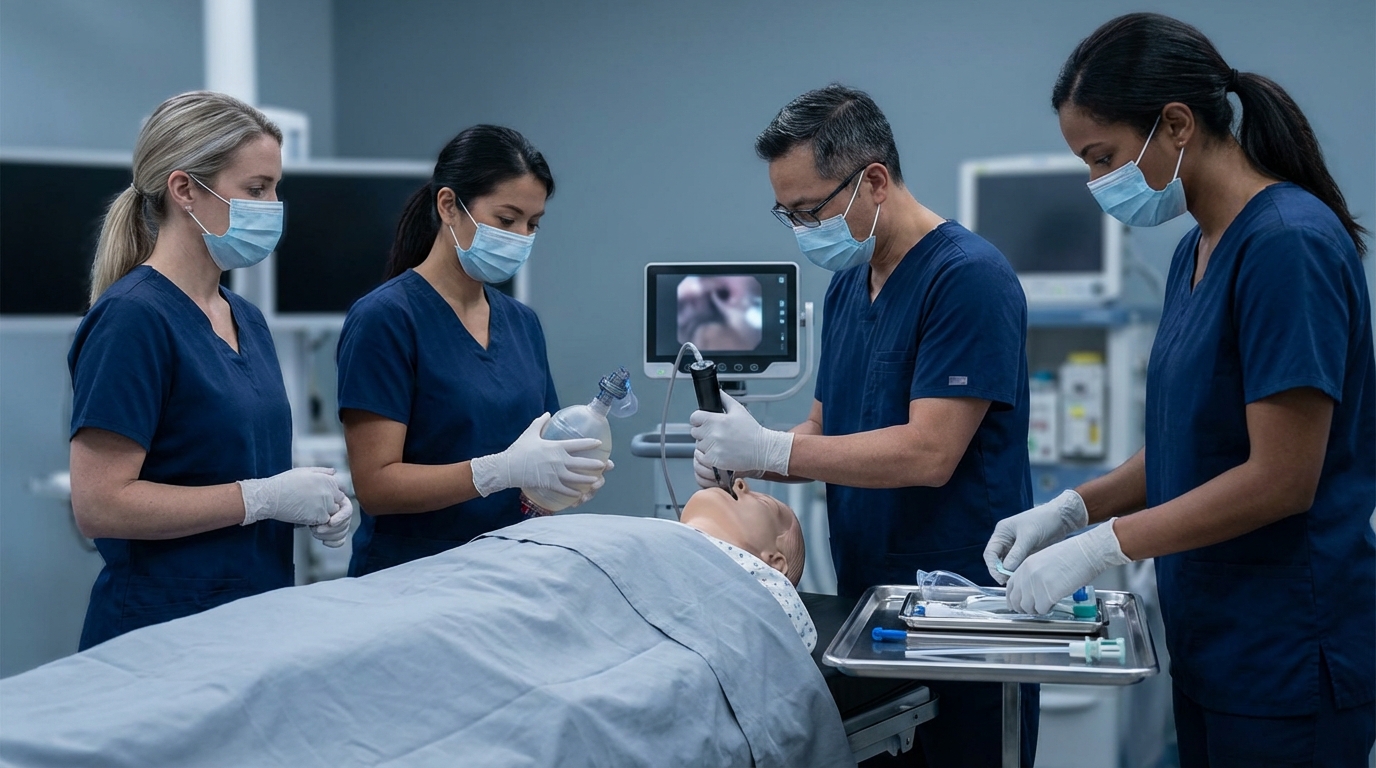
Practical Training
Airway management is a perishable skill – a competency that deteriorates without regular training. Rare but life-saving procedures such as emergency cricothyrotomy, CICO management, and structured RSI cannot be adequately practiced in routine clinical work. In the Emergency Physician Refresher course by Simulation Tirol, you train these scenarios hands-on using simulators and models, work through the airway algorithm under realistic time pressure, and sharpen your decision-making in critical moments. Because when the real situation occurs, what counts is not what you once read – but what you have trained.
More Articles
Acute Dyspnea: Differential Diagnosis and Immediate Management
Dyspnea has numerous causes ranging from asthma to heart failure to pulmonary embolism. This article presents a systematic approach to differentiation based on history, clinical examination, auscultation, and point-of-care diagnostics.
Acute Hypoglycemia: Emergency Management in Adults and Children
Threshold values, symptom recognition, oral glucose vs. IV dextrose vs. glucagon – with separate dosing for adult and pediatric patients. A common emergency presentation systematically reviewed.
Acute Adrenal Insufficiency: Addisonian Crisis in the Emergency Setting
Adrenal crisis is frequently misdiagnosed and can be fatal. This article describes at-risk patients, clinical signs, immediate hydrocortisone administration, and the management of accompanying hypoglycemia and hyperkalemia.