Mechanical CPR Devices: LUCAS and AutoPulse in Action
When are mechanical CPR devices indicated? Evidence, contraindications, correct application, and special situations such as transport CPR and the cardiac catheterization lab.

Author: Dr. med. univ. Daniel Pehböck, DESA
Specialist in Anesthesiology and Intensive Care Medicine, AHA-certified ACLS/PALS Instructor, Course Director Simulation Tirol
Reading time approx. 8 min
Mechanical CPR devices have become an established component of many EMS and hospital systems in recent years. Devices such as LUCAS (Lund University Cardiopulmonary Assist System) and AutoPulse promise consistent, fatigue-free chest compressions – an advantage that is particularly valuable in situations where manual CPR reaches its limits. Nevertheless, the evidence shows that mechanical CPR devices do not fundamentally replace manual resuscitation but rather serve as a meaningful supplement under defined conditions. This article examines the mechanism of action, indications, contraindications, correct application, and the current evidence – with a special focus on transport CPR, the cardiac catheterization lab, and other special situations.
Mechanism of Action: LUCAS vs. AutoPulse
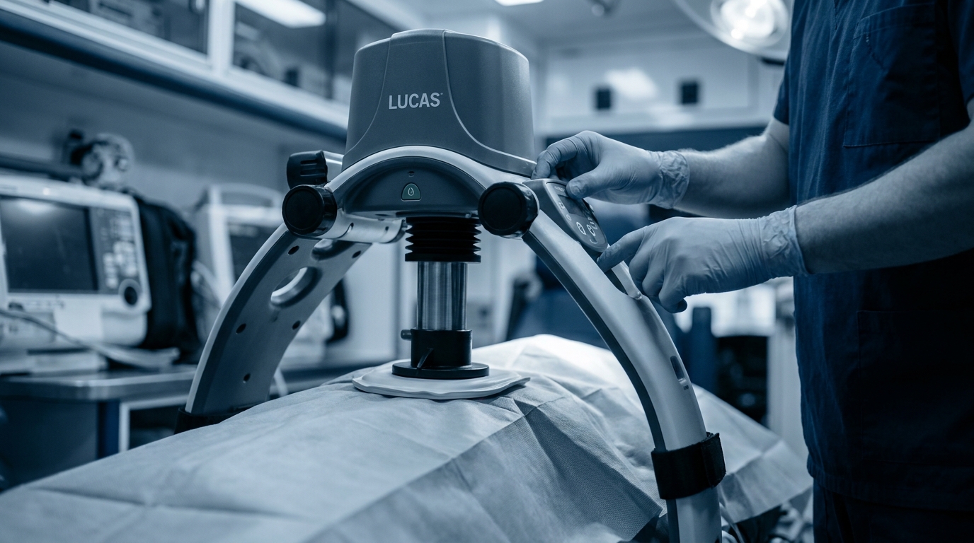
LUCAS
The LUCAS uses a piston-based compression mechanism. A suction-cup-like plunger is positioned over the lower third of the sternum and compresses the chest to a defined depth (approximately 53 mm in adults). Between compressions, active decompression ensures complete chest wall recoil. Depending on the generation, the LUCAS can be operated with an integrated battery or via an external compressed air supply. The compression rate is fixed at 102/min.
Key technical features:
- Compression depth: approximately 53 mm (± 2 mm)
- Rate: 102 compressions/min
- Active decompression via suction cup
- 30:2 mode (with ventilation pauses) and continuous mode available
- Weight approximately 7–8 kg depending on version
AutoPulse
The AutoPulse follows a fundamentally different concept. Instead of a piston, it uses a circumferential load-distributing band (LDB) that encircles the entire anterior thorax and compresses it through contraction. This distributes the compression force over a larger area. The device automatically calculates the optimal compression depth based on an initial chest circumference measurement (approximately 20% of the anterior-posterior chest diameter).
Key technical features:
- Circumferential compression via load-distributing band
- Automatic calibration to patient anatomy
- Compression rate: 80 compressions/min
- Platform weight: approximately 12 kg
- No active decompression
Comparison at a Glance
| Feature | LUCAS | AutoPulse |
|---|---|---|
| Compression mechanism | Piston/plunger | Load-distributing band |
| Rate | 102/min | 80/min |
| Active decompression | Yes | No |
| Calibration | Fixed setting | Automatic (chest circumference) |
| Ventilation pause mode | Yes (30:2) | Yes |
| Weight | ~7–8 kg | ~12 kg |
Evidence
The question of whether mechanical CPR devices improve outcomes compared to manual resuscitation has been the subject of intensive research. The data allow for differentiated conclusions.
Large Randomized Trials
Three large randomized controlled trials (RCTs) have significantly shaped the discussion:
- LINC Trial (LUCAS vs. manual CPR): This multicenter study showed no significant difference in 4-hour survival. A per-protocol analysis suggested a possible benefit of LUCAS, but this was not defined as the primary endpoint.
- PARAMEDIC Trial (LUCAS vs. manual CPR): Again, no survival benefit was found for mechanical CPR. There was even a trend toward worse neurological outcomes in the LUCAS group, which may be explained by longer no-flow times during device application.
- CIRC Trial (AutoPulse vs. manual CPR): This study demonstrated equivalence of both methods regarding survival to hospital discharge.
What the AHA Guidelines Say
The current AHA guidelines state the following recommendation: Mechanical CPR devices may be considered as a reasonable alternative to manual CPR when high-quality manual CPR is not feasible or places the provider at risk (Class IIb, Level of Evidence B-R). A general superiority over correctly performed manual CPR has not been demonstrated.
Interpretation for Clinical Practice
The trials have an important limitation: They primarily reflect standard prehospital use. In special situations – prolonged resuscitation, transport under CPR, cardiac catheterization lab – comparably large RCTs do not exist. However, these are precisely the situations where mechanical devices demonstrate their actual strengths, as registry data and cohort studies suggest.
Indications: When Mechanical CPR Makes Sense
Mechanical CPR devices are most beneficial where the quality of manual CPR is predictably compromised.
Primary Indications
- Transport CPR: In a moving ambulance, helicopter, or during intrahospital transfer, high-quality manual CPR is virtually impossible. Studies show that compression depth and rate decrease significantly under transport conditions. Mechanical devices provide consistent compression quality in these scenarios.
- Prolonged resuscitation: When a long resuscitation duration is anticipated (e.g., hypothermia, intoxication, eCPR preparation), rescuers fatigue despite switching every two minutes. Mechanical devices maintain compression quality for hours.
- Cardiac catheterization lab (cath lab): Under the fluoroscopy C-arm, manual CPR is problematic due to radiation exposure to staff and limited space. Mechanical devices enable continuous CPR during percutaneous coronary intervention (PCI).
- Preparation for eCPR (ECMO-CPR): During cannulation for extracorporeal CPR, mechanical devices provide reliable circulatory support without the need to dedicate personnel to compressions.
- Limited personnel resources: In mass casualty incidents or other situations with staffing shortages, mechanical devices can "free up" a team member.
Relative Indications
- Resuscitation in confined spaces (e.g., stairwells, small apartments)
- Organ donation after circulatory death (DCD – Donation after Circulatory Death): maintaining organ perfusion
- Resuscitation in morbidly obese patients (rescuer fatigue occurs more rapidly)
Contraindications
Not every patient is suitable for mechanical CPR. The following contraindications should be considered:
Absolute contraindications:
- Children and infants (devices are designed and validated for adults)
- Traumatic cardiac arrest with massive chest wall instability
- Patients whose body habitus does not fit device specifications (too small, too tall, chest too wide)
Relative contraindications:
- Known severe aortic pathology (e.g., type A aortic dissection – though the overall situation must be evaluated)
- Thoracic deformities that prevent correct positioning
- Implanted LVAD systems (case-by-case decision)
With the LUCAS, the plunger must be positioned exactly over the lower third of the sternum. With the AutoPulse, the band must be able to fully encircle the chest. If the patient's anatomy does not fit the device, manual CPR is the better choice.
Correct Application: Step by Step
Applying a mechanical CPR device must not significantly prolong hands-off time. The AHA emphasizes that interruptions in chest compressions must be reduced to an absolute minimum.
LUCAS – Application Sequence
- Preparation: Have the device ready, check battery. Have the backplate ready.
- Continue manual CPR: At no point interrupt compressions longer than necessary.
- Position the backplate: During the next planned compression pause (e.g., rhythm check), slide the backplate under the patient. This is best achieved by slightly elevating the upper body or log-rolling the patient.
- Mount the compression unit: Click the piston unit into the backplate. Position the plunger over the lower third of the sternum.
- Adjust height: Lower the plunger to the chest surface.
- Start: Select the mode (30:2 or continuous) and activate the device.
- Verify position: Confirm correct plunger position visually and, if possible, with etCO₂ monitoring.
Time target: The entire application procedure should be achievable in under 20 seconds (ideally under 10–15 seconds) without compression pauses when practiced as a team.
AutoPulse – Application Sequence
- Position the platform: Place the platform next to the patient and position the upper body onto it (during the next planned pause).
- Apply the band: Pull the load-distributing band over the anterior chest and click it into place.
- Calibration: The device performs an automatic calibration measurement.
- Start: Activate compression mode.
Common Errors During Application
- Excessively long hands-off time due to lack of team communication
- Incorrect plunger position (too cranial → ineffective compressions, risk of injury)
- Forgetting to recalibrate the device after repositioning (AutoPulse)
- Continuing resuscitation without etCO₂ verification after device application
Special Situations in Detail
Transport CPR
Transport CPR is probably the most widely accepted indication for mechanical CPR. In a moving ambulance, studies using accelerometer measurements show that manual compressions decrease massively in depth and rate – sometimes by 30–50% compared to stationary conditions. Additionally, rescuer safety in a moving vehicle during manual CPR is compromised.
Recommendations for transport CPR:
- Apply the mechanical device before initiating transport if possible
- Transport only when clearly indicated (e.g., for PCI, eCPR, rewarming)
- Continuous etCO₂ monitoring for quality control throughout transport
- Regular rhythm checks even under mechanical CPR
Cardiac Catheterization Lab
In the cath lab, mechanical CPR enables emergency PCI in patients with refractory cardiac arrest. The LUCAS is often more compatible with C-arm positioning due to its lower profile compared to the AutoPulse. Registry data show that patients with STEMI-related cardiac arrest who receive successful PCI under mechanical CPR can achieve neurologically intact survival rates of 15–25%.
Practical considerations in the cath lab:
- Clarify beforehand whether the device is compatible with the local C-arm
- Keep the femoral catheter access site clear
- With LUCAS: verify plunger position again before fluoroscopy
Hypothermic Resuscitation
In accidental hypothermia with cardiac arrest, the principle applies: "Nobody is dead until warm and dead." Resuscitation may continue for hours until extracorporeal rewarming is initiated. Mechanical CPR is virtually indispensable here to maintain adequate perfusion throughout the entire period.
eCPR Bridging
In refractory cardiac arrest with a reversible cause, extracorporeal CPR is gaining increasing importance. Cannulation takes 15–30 minutes even in experienced centers. Mechanical CPR during the cannulation phase standardizes circulatory support and reduces the burden on the team.
Quality Control During Mechanical CPR
Even mechanical devices do not replace clinical monitoring. The following parameters should be continuously monitored:
- End-tidal CO₂ (etCO₂): The most important surrogate parameter for compression effectiveness. A sudden increase may indicate ROSC. Values persistently below 10 mmHg suggest ineffective CPR – even under mechanical compression.
- Arterial pressure (if invasively monitored): Diastolic pressure > 25 mmHg as a target for coronary perfusion.
- Clinical signs: Pupil reaction, skin color, spontaneous movements.
- Device function: Battery charge, band integrity (AutoPulse), plunger position (LUCAS).
Team Training and CRM Aspects
Integrating mechanical CPR devices into the resuscitation algorithm requires team training. The most common errors arise not from device failure but from:
- Lack of role assignment (Who retrieves the device? Who applies it?)
- Prolonged hands-off times with unpracticed application
- Fixation on the device during technical problems instead of reverting to manual CPR
- Failure to recalibrate after patient repositioning
A clear team briefing before deployment and regular simulation training significantly reduce these error sources.
Summary of Key Messages
- Mechanical CPR devices are not superior to correctly performed manual CPR in standard situations.
- Their primary benefit lies in special situations: transport CPR, cath lab, prolonged resuscitation, eCPR bridging, hypothermia.
- Application must be trained as a team to minimize hands-off times.
- etCO₂ monitoring is essential even during mechanical CPR.
- Observe contraindications (children, unsuitable anatomy, severe chest wall instability).
- When in doubt: Manual CPR remains the standard – mechanical devices are a tool, not a replacement for structured teamwork.
Practical Training
The correct integration of mechanical CPR devices into the ACLS algorithm is best trained under realistic conditions. In the AHA-certified ACLS courses offered by Simulation Tirol, you practice structured resuscitation as a team – from basic management to special situations where you must make decisions about device selection and application under time pressure. Because even the best equipment is only as effective as the team using it. You can find all information about ACLS Provider and Refresher courses on the Simulation Tirol website.
Want to practice this hands-on?
In our ACLS-Kurs (Advanced Cardiac Life Support) you practice this topic hands-on with high-tech simulators and experienced instructors.
More Articles
Acute Heart Failure: Emergency Management and Differentiation
From pulmonary edema to cardiogenic shock – acute heart failure requires rapid hemodynamic classification. This article explains the clinical classification and first-line therapy with nitrates, diuretics, and vasopressors.
Acute Coronary Syndrome: Initial Management and the MONA Approach
ACS is one of the most common cardiac emergencies. This article covers initial diagnostics (12-lead ECG, troponin), risk stratification, pharmacological first-line treatment, and decision pathways for reperfusion therapy.
ECG Rhythm Interpretation: The Most Important Emergency Rhythms
From ventricular fibrillation to pulseless electrical activity to third-degree AV block – this article presents typical ECG patterns of the most critical emergency cardiac arrhythmias with recognition criteria and immediate therapy.